Skin Check
Regular annual skin check is recommended for Queenslanders with fair skin and higher risk of skin cancer. Skin cancer is the most common cancer in the Australia; in fact, 1 in 3 Australians will develop skin cancer in their lifetime. People of all colours and races can get skin cancer. There are many different types of skin cancer, including basal cell carcinoma (BCC), squamous cell carcinoma (SCC), and melanoma.
BCC and SCC are the most common forms of skin cancer, but melanoma is the most deadly.
With early detection and proper treatment, the cure rate for BCC and SCC is about 95 percent. When melanoma is detected before it spreads, it has a high cure rate. Regular self-skin exams and a yearly examination by a skin cancer specialist help people find early skin cancers.
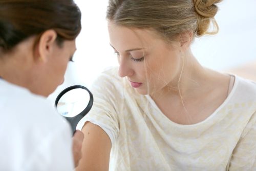
Skin cancer doctors use tools and techniques to check skin thoroughly, beyond what the naked eye can see. It is important to get a professional skin check by a doctor who is trained in diagnosing and management of skin cancers. QLD Skin Cancer Clinic offers the latest technology for examining skin spots.

What to expect at your skin check?
When you arrive at QLD Skin Cancer Clinic you will be asked to complete a patient questionnaire. Please bring a list of your current medications as well as your local GP details to help complete your details. During your consultation, doctor will assess your medical and family history, particularly in relation to previous skin cancers, medications, occupation and whether you are allergic to medications.
Your doctor will also want to know whether you take medications that thin the blood (eg Aspirin, Warfarin or Plavix) or whether you have a pacemaker. Please mention any particular spots that you are concerned about to your doctor.

What skin cancer screening technology do you use?
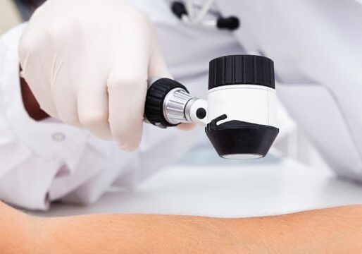
We have high-magnification digital camera and accessories for monitoring skin spots. Doctor usually uses a high resolution screen to analyse the the spot to get extra information about the likelihood of melanoma.
Doctor may take photographs of your whole body, or regions of your body. If these photographs are repeated, we can automatically determine on your next visit if you have developed new spots or if there are significant changes in the spots that were already present. This procedure, known as total body photography, is most useful for people with too many moles.
Your doctor will offer total body mapping if he or she thinks it would be useful, but in most cases a thorough full body examination with the dermatoscope is sufficient. If you would like total body mapping and your doctor hasn’t mentioned it, please feel free to ask about it.
Your doctor will show you the photographs and point out any suspicious features. This will help you and the doctor decide if any of the spots must be treated soon, or if they should be observed for changes over a period of time.
Checking suspicious lesions
If you have a suspicious skin lesion on your body, doctor may recommend to take a sample (skin biopsy) for further analysis of the lesion at histopathology lab.
Some melanomas grow very slowly and in the early stages may not show any obvious clues to their malignancy, even on close and careful examination using a dermatoscope. In rare cases such as these, they may be diagnosed by monitoring an unusual ‘mole’ over time. This involves taking a digital photograph of the lesion with a magnified view through the dermatoscope, and then comparing the original image with a follow-up one taken at a later appointment. This is usually a 3 or 6 month interval, depending on the lesion.
If you are diagnosed with a skin cancer, your doctor will be able to work out a treatment plan that is best for you. Most skin cancers can be treated in the purpose built rooms at the centre, either with surgery or occasionally a non-surgical treatment.
These procedures are commonly performed during your consultation if time allows: – Cryotherapy (freezing) of solar keratoses, warts and some other lesions – Electrosurgery (burning) of sunspots or angiomas (small spots or lumps composed of blood vessels) – Simple shave biopsies of suspicious lesions not requiring stitches. You would normally need to return at a later date for more complicated and time-consuming procedures (for example, requiring stitches).


